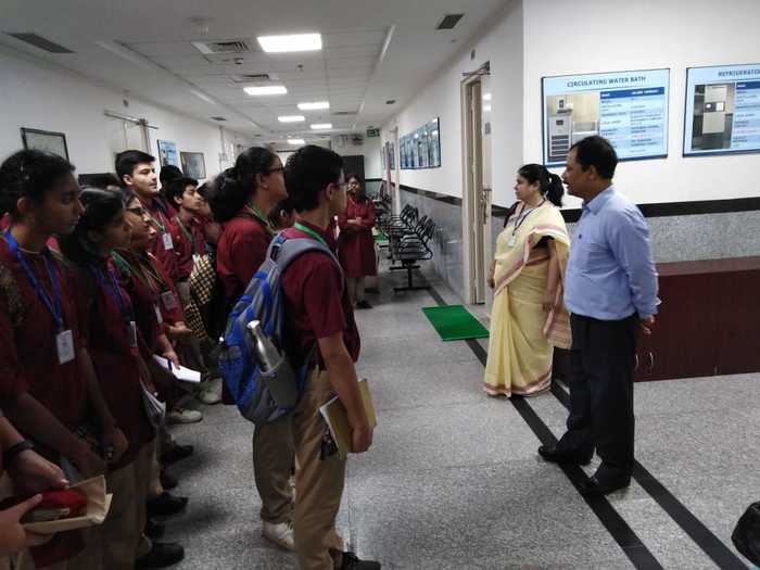
Visit to view the Electron Microscope Facility at AIIMS
Students of class IX had the elevating experience of visiting the Electron Microscope Facility at AIIMS on 17th, 20th and 21st May 2019. 105 students visited the Facility in groups of 3 accompanied by 2 teachers. Each group comprised 35 students.
Students had the opportunity to meet a very learned and venerated person, Dr TC Nag, Professor of Anatomy at AIIMS, who explained the concept of Microscopes in detail to all the students. He discussed the difference between resolution and working of a Light and Electron Microscope. He also clarified the students doubts in a very patient and detailed manner.
[gallery link="file" columns="3"]
This was followed by a visit to the Ultra microtome Machine Room where students could learn how thin slides for observation were prepared using an ultra-microtome. Next in line was a visit to see the Scanning Electron Microscope (SEM) where students were shown specimens observed under SEM along with pictures of the results on a computer screen. It included various structures such as a hair strand, cat fish spikes etc. The most striking feature of the session was the Transmission Electron Microscope (TEM). It was huge in size with an injection slot where the sample to be studied was injected. It had a vacuum chamber for electrons to travel and a phosphorus sheet to take the impressions. The images of various cells, tissues and cell organelles obtained through the electron microscope were shown to the students, which added to the excitement and enthusiasm of us all!
It was an exhausting trip but definitely worth the fatigue. The students enjoyed it to the fullest and came back with lots of practical learning and knowledge