An enlightening visit to the Institute of Genomics and Integrated Biology (CSIR), Bio students of classes 11 and 12.
Science is the poetry of reality.
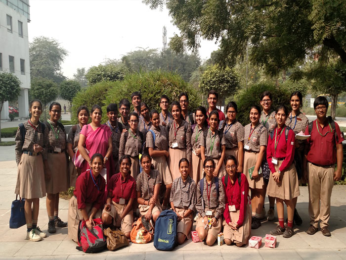
Twenty seven Biology students of classes 11 and 12 participated in an Open Day session at the Institute of Genomics and Integrative Biology (IGIB), a premier Institute of the Council of Scientific and Industrial Research (CSIR) on 4th November 2016. They were accompanied by their Biology teacher, Ms. Namrata Jit Kaur.
IGIB engages in research of national importance in the areas of genomics, molecular medicine, bioinformatics, proteomics and environmental biotechnology. Several groups at the IGIB are also involved in studying the molecular basis of human diseases especially neuropsychiatric disorders such as schizophrenia, cardiovascular diseases and diabetes. A significant number of IGIB scientists also focus on respiratory diseases using clinical, genetic, molecular and drug development approaches to tackle this challenging area. IGIB has over the years built up expertise in high-throughput data analysis and genome annotation. Scientists at IGIB are also exploring the rich microbial diversity of India and developing biotechnological applications using this resource to address issues pertaining to the environment and energy crisis. Ayurgenomics is such an emerging inter-disciplinary project that draws upon the methodology and rigor of modern genomics and the traditional knowledge base of Ayurveda.
[gallery link="file"]
Students were first shown short interesting clips in the auditorium. They were explained several concepts with simplicity and attractive illustrations, stretching from the process of our cell making antibodies to the RNA world hypothesis!
The students were divided into two batches and shown around the institute by tour guides who encouraged students to participate by handing certificates to the most interactive ones!
The students were taken to an electron microscope lab where they got exposure to the concept of microscopy. An electron microscope is one with high magnification and resolution, employing electron beams in place of light and using electron lenses. There are two types among these, the transmission electron microscope and the scanning electron microscope. While the former provides two dimensional images of a sample from the inside, the latter gives a three dimensional image, providing morphological and topological details. We were shown the components and functioning of both the electron microscopes. While the resolution of the transmission electron microscope is 0.2nm, the resolution of the scanning electron microscope is about 10nm. We were also shown various images under the two microscopes: human cells and its various parts, yeast cells, embryo of zebra fish, a hair follicle and bundles of sarcomeres. It was extremely interesting to view such intricate structures of the microscopic world.
The most interesting room was THE CELL. The whole room resembled a 3 dimensional structure of an animal cell with the nucleus in the middle and the other cell organelles spread across the floor. First, we had a quiz about the cell. Next, we had an activity which required us to find the DNA in the cell, its transcribed mRNA, and then translate it into a polypeptide chain using beads. This was a fun filled and interactive session.
After this, we visited the exhibit area. One stall showed why and how cells age. The stall even had an interesting game which involved students looking at photos of people for a very short duration and then judging their ages. The next stall displayed various nanoparticles. We were even shown the preparation of nanoparticles of Ferrous Oxide.
Another eye catching exhibit was the chemiluminescence. For this display, we all were led into a completely dark room. All of us were mesmerised by the sight of tiny florescent lights hanging from the roof. A while later it was revealed that those tiny lights were actually chemiluminescent substances that glow in the dark. There we were also informed about phospholuminescence whose daily life examples are the clock dial, party sticks, AC remote controls, etc.
Our next hands'-on-experience was experimenting with our own saliva. Saliva was taken in a small microfuge tube, SDS (sodium dodecyl sulphate) chemical and ethanol was added to it and then was allowed to precipitate for 3 minutes. We were overwhelmed to see the white entangled strands of our DNA, which were isolated and extracted from our cells, with our naked eyes. We were overjoyed when we were told that we could carry our DNA home!
The students were then shown equipment used by the scientists at IGIB. We were shown how incubators work. They provide controlled environment for bacteria cultures. We were even shown the Laminar flow cabinet. It is a carefully enclosed bench designed to prevent contamination of semiconductor wafers, biological samples and other particle sensitive materials.
Lastly, we saw the huge supercomputer which stores all the data of the institute. It has firewall protection to prevent hacking. The supercomputer is stored in a cool area to absorb the heat produced and the area is protected well to prevent unauthorised access. We were even shown a shelf of the supercomputer to give us an idea of its components.
We thoroughly enjoyed visiting this institute and would love to visit it again. This trip not only helped us in learning new facts but also boosted our enthusiasm towards science. After all,
The science of today is the technology of tomorrow.
Saruby, Muskan, Kara , Azaz, Priyadharshini, Yashmayi ( XI & XII Biology Students).